Brain aneurysms, which affect as many as one in 50 people, occur when a blood vessel wall weakens and bulges, setting the scene for a potentially deadly rupture. Now scientists have created a 3-D-printed aneurysm model in the laboratory and “operated” on it: they inserted a device to seal it off and prevent it from bursting. Such models could be tailored to replicate an individual patient's blood vessel, letting doctors try different treatments and find the best solution.
To treat an aneurysm, brain surgeons sometimes operate to install a metal clip on the ballooning vessel that prevents the pooling of blood. A less invasive method involves inserting tiny metal coils into the aneurysm via a catheter to induce a blood clot that seals it off. Most treatment devices are tested in animals, whose blood vessels do not perfectly resemble those in humans. And previous lab-dish aneurysms could not mimic the properties of living blood vessels. “We thought maybe there could be a better way of testing those [treatment] devices,” says Lindy Jang, a biomedical engineering graduate student at Texas A&M University, who led the new study, published in Biofabrication.
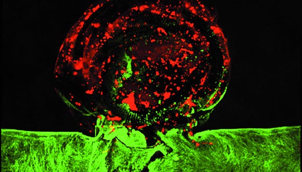
Jang and her colleagues 3-D-printed an aneurysm structure with a water-based gel and populated it with human cells that line the brain's blood vessels. They then operated on the aneurysm, injecting platinum coils into the bulging vessel. Finally, they filled the blood vessel with plasma (the liquid component of blood), which formed a clot that sealed off the bulge.
“We're trying to streamline the treatment of aneurysms and take the guesswork out,” says William “Rick” Hynes, a study co-author and biofabrication research engineer at Lawrence Livermore National Laboratory who performed the surgery. “The goal is to use these devices to validate models so someone could take a 3-D scan, recreate it in the simulation, then try adding [blood] flow and determine if they need to treat the aneurysm or leave it alone.”
“I think it's really significant,” Matthew Gounis, a biomedical engineer at the University of Massachusetts Medical School, says of the new model. Other groups have developed aneurysm models, but this one is exciting because it better replicates a human blood vessel by adding living cells, says Gounis, who was not involved in the new study. Surgeons could practice on such models before operating on a real patient, he says: “If you have a particularly challenging case, you can print out the case, and you can basically practice before you get to the patient, in their anatomy.”
 Rights & Permissions

