
In my first neuroscience course at Columbia University, I learned about the homunculus. This “little man” is depicted as an upside-down representation of the human body moving from toe to head in a portion of the cerebral cortex that controls movement. Wilder Penfield, the trailblazing Canadian-American neurosurgeon, created the homunculus metaphor after mapping areas of the human brain by using direct electrical stimulation in awake patients in the 1930s.
From this work, a nurse working with Penfield created one of science’s most iconic illustrations. It shows the homunculus stretched out over the brain’s surface, a small body with enlarged mouth, hands and feet, each part exaggerated in proportion to the amount of neural real estate occupied. Later three-dimensional representations of the homunculus depict it as a grotesque, hairless goblin with enormous lips, hands and feet. The somewhat frightening creature is prominent in every neuroscience textbook and even has its own Dungeons & Dragons character, with bat wings for good measure.
As a student and later as a practitioner in the field, I had accepted the homunculus as unquestioned fact. As a professor, I would dutifully teach students that the distorted figure represented a key aspect of brain organization. The evidence seemed overwhelming: stimulation of brain areas that corresponded to the relevant areas of the motor cortex, which generates signals to direct body movement, elicited the expected foot, hand and face muscle twitches. The neuroimaging techniques similarly map movement of a hand or toe to the matching brain area, and damage from a stroke impedes movement in the expected body part. Given all this, any new research of this brain area, seemed like a nonstarter. It had been definitively mapped 90 years ago, So, I focused my own research elsewhere.
What ultimately made our team go back to replicate Penfield’s original work was a set of anomalous findings using a specialized type of MRI that identifies brain networks dedicated to a particular function. Such “resting state” functional neuroimaging looks for spontaneous activity that occurs simultaneously in different regions while a person is lying at rest in a scanner.
About three years ago, results from these latter-day imaging methods clashed violently with the classical neuroanatomy from Penfield’s era. The problem started when I was trying to decide how much to trust a new method for cleaning up imaging data and had asked to conduct what I thought was a routine test of its validity by checking the connections in the area of the homunculus that corresponded to the hand.
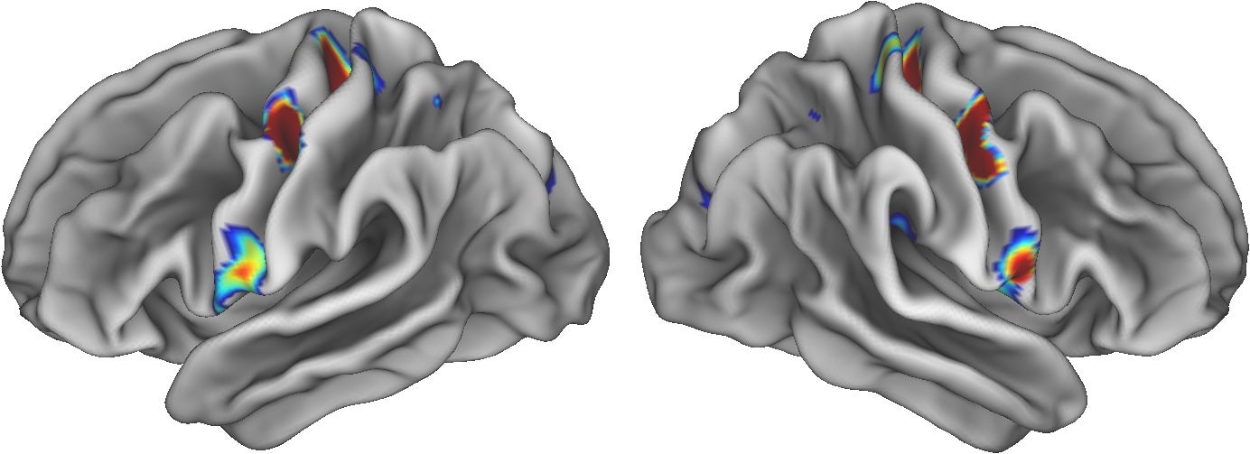
The homunculus’ hand showed the expected connections to the hand region in the other half of the brain, as did the relevant areas of the motor cortex that control movement of the foot and mouth. But when we moved up or down this brain section that makes up the homunculus, we were in for a surprise.
A seemingly absurd pattern popped out in the imaging that showed three interconnected and previously undocumented areas right in the homunculus section of the motor cortex. Motor homunculus regions were thought to only be linked to each other across the brain’s hemispheres, the left hand with the right hand, and the left foot with the right foot. Connections to varying locations within the same hemisphere were not supposed to exist. I tried to dismiss this weird looking “three-spot” imaging motif as an error, but in discussing it we also acknowledged the remote possibility that what we had seen might somehow contradict the classical neuroscience supporting a homunculus. We continued to explore by trying different methods.
I could not make sense of the result. Why would there be this separate fundamentally different profile of brain connections right in the middle of this section of motor cortex if there was only a single integral homunculus? What was this distinct brain network right in the middle of the homunculus? I filed the broken homunculus discovery in the “does not compute” folder of my brain and tried to work on other projects. But our findings kept bugging me.
At one point, my collaborator Evan Gordon and I were examining data that revealed that the mystifying set of three spots in the homunculus were connected to a cognitive control network important for planning future actions, which I had been studying since graduate school. These connections to higher-order control regions planted the idea that the three-spot network might be important for merging motor command signals from the homunculus with neural activity for more abstract planning.
If that were the case, it implied that maybe, just maybe, the classic “little man” figure of the homunculus needed to be redrawn. Evan and I made a pact to chase this conflicting finding wherever it might lead, even if it seemed to challenge the explanations found in neuroscience textbooks. We went on to reexamine our own prior work, pulled in all potentially relevant data sets we could get our hands on, conducted new experiments and reexamined the published evidence for and against the homunculus story, going back to the early 1900s.
The quest to solve the mystery of the three spots was filled with many surprises. Early on, Evan excitedly announced that our own prior publications had already revealed the location of the spot triad. I was incredulous and fixated on these three imaging blemishes, I had spent so much time with the data that they infiltrated my dreams.
As part of our quest, we became aware of contradictory findings in macaques and other nonhuman primates, which had not at the time sufficed to challenge the homunculus metaphor. Digging through Penfield’s original brain stimulation data from about 90 years ago revealed that these conflicting results were equally, if not more, consistent with other models of motor cortex organization that did not conform to the homunculus metaphor.
Looking still further, we discovered that our findings could be reconciled with other relatively recent research. In 2002, Michael Graziano, a Princeton University neuroscientist, and colleagues had found that the same area of the motor cortex is responsible in nonhuman primates for controlling feeding, defensive behaviors and other actions more complex than movement of, say, the legs or lips. This and other research began to nudge us toward the conclusion that, after 90 years, the homunculus model of the homunculus was ready for retirement. The idea of a region of motor cortex that roughly mirrored the head-to-toe alignment of the human body could no longer prevail.
We put forward evidence for our views in a recent Nature article. Instead of a continuous head-to-toe representation of body movements in a single homunculus, our findings show this neural representation of the body is sliced into three sections, one for the feet, another for the hands and a third for the mouth. Separating or adjoining these areas are the locations of the mysterious three spots that produced so much frustration for us. All three turn out to be interconnected, and we found that they are responsible for a range of tasks, including planning, regulating internal organs and even becoming active when someone is just thinking about a making some movement—in essence, forming a link between mind and body.
This network, which we named the Somato-Cognitive Action Network (SCAN), executes a plan to move the whole body. It integrates mind and body by linking to other brain regions controlling breathing heart rate, muscle tension, even butterflies in the stomach, all of which provide feedback for planning future actions—here’s what is needed to avoid that upset stomach or laceration next time. SCAN also connects to regions important for drive and motivation, damage to which can induce apathy. The recognition that body control and motor activity are represented by a common brain circuit helps explain why mind and body states so often interact.
The positive effects of exercise or electrical stimulation of the motor cortex to relieve chronic pain start to make sense upon the realization that pain and whole-body movements are controlled by the same brain network. If arousal and initiating some physical actions are part of the same network, it also seems less paradoxical to treat difficulties of staying on task and hyperactivity with stimulants.
Overall, our findings mean that the homunculus wears no clothes. How could I have missed for so long what was so obvious? It has been eye-opening to realize how powerfully my thinking is shaped by prior assumptions. My trust in textbook teachings blinded me to data that were right there in front of us. As time went on, our confidence in our data grew to the point that we eventually trusted it more than the prevailing neuroscience doctrine.
Good stories are powerful, even in the nominally objective realm of science. The homunculus likely lived to the age of 90 because everyone loves a good story. The imagery of the distorted figure of the homunculus, with outsized lips and hands, was so compelling that it took on a life of its own. In his book Penfield highlighted that the homunculus was mainly a model for teaching medical students; that it must not be overinterpreted. That warning never made it into the textbooks.
This is an opinion and analysis article, and the views expressed by the author or authors are not necessarily those of Scientific American.
 Rights & Permissions
