Networks pervade our lives. Every day we use intricate networks of roads, railways, maritime routes and skyways traversed by commercial flights. They exist even beyond our immediate experience. Think of the World Wide Web, the power grid and the universe, of which the Milky Way is an infinitesimal node in a seemingly boundless network of galaxies. Few such systems of interacting connections, however, match the complexity of the one underneath our skull.
Neuroscience has gained a higher profile in recent years, as many people have grown familiar with splashily colored images that show brain regions “lighting up” during a mental task. There is, for instance, the temporal lobe, the area by your ear, which is involved with memory, and the occipital lobe at the back of your head, which dedicates itself to vision.
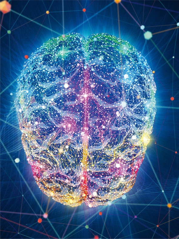
What has been missing from this account of human brain function is how all these distinct regions interact to give rise to who we are. Our laboratory and others have borrowed a language from a branch of mathematics called graph theory that allows us to parse, probe and predict complex interactions of the brain that bridge the seemingly vast gap between frenzied neural electrical activity and an array of cognitive tasks—sensing, remembering, making decisions, learning a new skill and initiating movement. This new field of network neuroscience builds on and reinforces the idea that certain regions of the brain carry out defined activities. In the most fundamental sense, what the brain is—and thus who we are as conscious beings—is, in fact, defined by a sprawling network of 100 billion neurons with at least 100 trillion connecting points, or synapses.
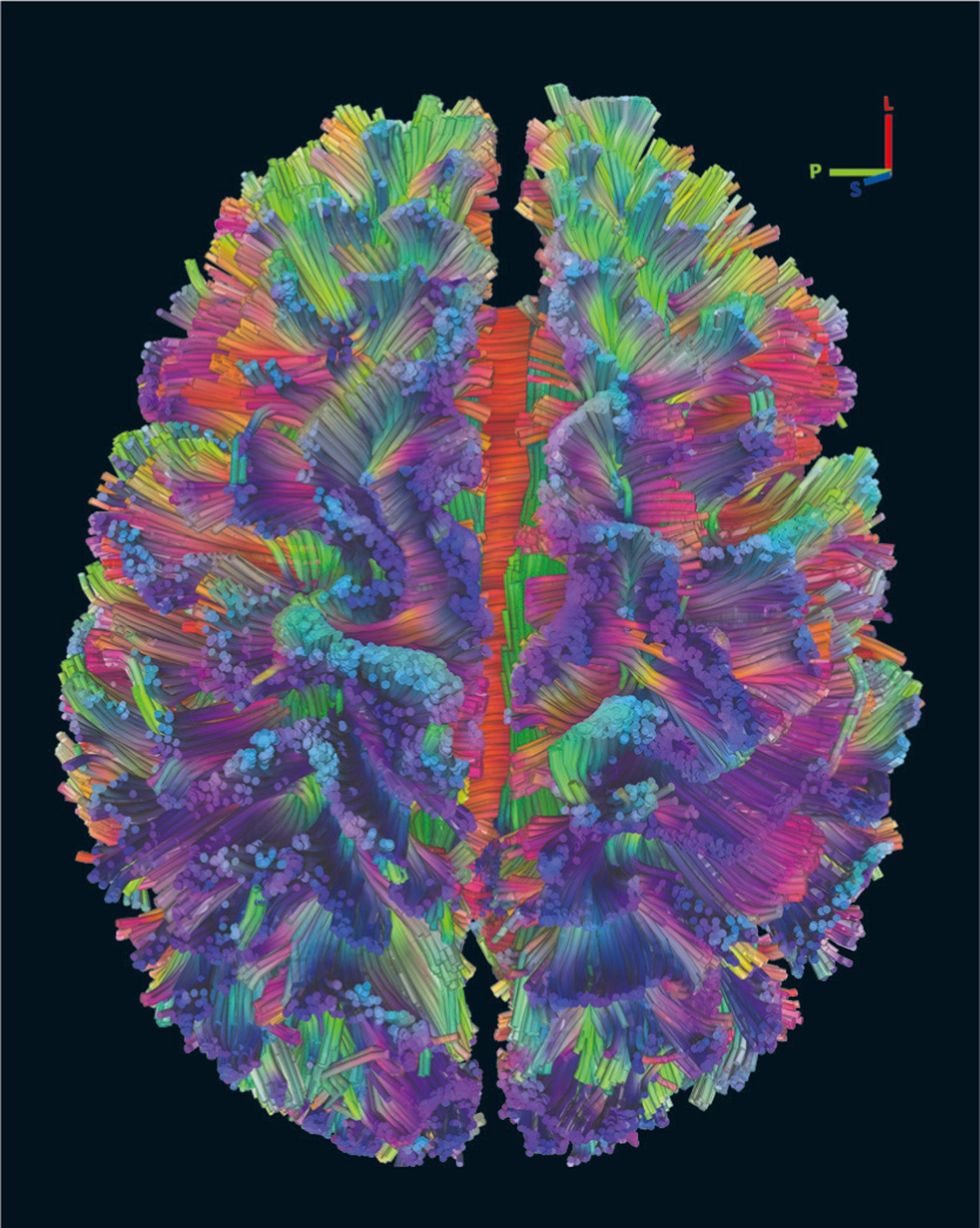
Network neuroscience seeks to capture this complexity. We can now model the data supplied by brain imaging as a graph composed of nodes and edges. In a graph, nodes represent the units of the network, such as neurons or, in another context, airports. Edges serve as the connections between nodes—think of one neuron intertwined with the next or contemplate airline flight routes. In our work, the human brain is reduced to a graph of roughly 300 nodes. Diverse areas can be linked together by edges representing the brain’s structural connections: thick bundles of tubular wires called white matter tracts that tie together brain regions. This depiction of the brain as a unified network has already furnished a clearer picture of cognitive functioning, along with the practical benefit of enabling better diagnoses and treatment of psychiatric disorders. As we glimpse ahead, an understanding of brain networks may lead to a blueprint for improved artificial intelligence, new medicines and electrical-stimulation technology to alter malfunctioning neural circuitry in depression—and perhaps even the development of genetic therapies to treat mental illness.
The Music of the Mind
To understand how networks underlie our cognitive capabilities, first consider the analogy of an orchestra playing a symphony. Until recently, neuroscientists have largely studied the functioning of individual brain regions in isolation, the neural equivalent of separate brass, percussion, string and woodwind sections. In the brain, this stratification represents an approach that dates back to Plato—quite simply, it entails carving nature at the joints and then studying the individual components that remain.
Just as it is useful to understand how the amygdala helps to process emotions, it is similarly vital to grasp how a violin produces high-pitched sounds. Still, even a complete list of brain regions and their functions—vision, motor, emotion, and so on—does not tell us how the brain really works. Nor does an inventory of instruments provide a recipe for Beethoven’s Eroica symphony.
Network neuroscientists have begun to tame these mysteries by examining the way each brain region is embedded in a larger network of such regions and by mapping the connections between regions to study how each is embedded in the large, integrated network that is the brain. There are two major approaches. First, examining structural connectivity captures the instrumentation of the brain’s orchestra. It is the physical means of creating the music, and the unique instrumentation of a given musical work constrains what can be played. Instrumentation matters, but it is not the music itself. Put another way, just as a collection of instruments is not music, an assemblage of wires does not represent brain function.
Second, living brains are massive orchestras of neurons that fire together in quite specific patterns. We hear a brain’s music by measuring the correlation between the activity of each pair of regions, indicating that they are working in concert. This measure of joint activity is known as functional connectivity, and we colloquially think of it as reflecting the music of the brain. If two regions fire with the same time-varying fluctuations, they are considered to be functionally connected. This music is just as important as the decibels produced by a French horn or viola. The volume of the brain’s music can be thought of as the level of activity of electrical signals buzzing about one brain area or another.
At any moment, though, some areas within the three-pound organ are more active than others. We have all heard the saying that people use a small fraction of their brain capacity. In fact, the entire brain is active at any point in time, but a given task modulates the activity of only a portion of the brain from its baseline level of activity.
That arrangement does not mean that you fulfill only half of your cognitive potential. In fact, if your entire brain were strongly active at the same time, it would be as if all the orchestra members were playing as loudly as possible—and that scenario would create chaos, not enable communication. The deafening sound would not convey the emotional overtones present in a great musical piece. It is the pitch, rhythms, tempo and strategic pauses that communicate information, both during a symphony and inside your head.
Modularity
Just as an orchestra can be divided into groups of instruments from different families, the brain can be separated into collections of nodes called modules—a description of localized networks. All brains are modular. Even the 302-neuron network of the nematode Caenorhabditis elegans has a modular structure. Nodes within a module share stronger connections to one another than to nodes in other modules.
Each module in the brain has a certain function, just as every family of instruments plays a role in the symphony. We recently performed an evaluation of a large number of independent studies—a meta-analysis—that included more than 10,000 functional magnetic resonance imaging (fMRI) experiments of subjects performing 83 different cognitive tasks and discovered that separate tasks map to different brain-network modules. There are modules occupied with attention, memory and introspective thought. Other modules, we found, are dedicated to hearing, motor movement and vision.
These sensory and motor cognitive processes involve single, contiguous modules, most of which are confined to one lobe of the brain. We also found that computations in modules do not spur more activity in other modules—a critical aspect of modular processing. Imagine a scenario in which every musician in an orchestra had to change the notes played every time another musician changed his or her notes. The orchestra would spiral out of control and would certainly not produce aesthetically pleasing sounds. Processing in the brain is similar—each module must be able to function mostly independently. Philosophers as early as Plato and as recent as Jerry Fodor have noted this necessity, and our research confirms it.
Even though brain modules are largely independent, a symphony requires that families of instruments be played in unison. Information generated by one module must eventually be integrated with other modules. Watching a movie with only a brain module for vision—without access to the one for emotions—would detract greatly from the experience.
For that reason, to complete many cognitive tasks, modules must often work together. A short-term memory task—holding a new phone number in your head—requires the cooperation of auditory, attention and memory-processing modules. To integrate and control the activity of multiple modules, the brain uses hubs—nodes where connections from the brain’s different modules meet.
Some key modules that control and integrate brain activity are less circumspect than others in their doings. Their connections extend globally to multiple brain lobes. The frontoparietal control module spans the frontal, parietal and temporal lobes. It developed relatively recently on the timescale of evolution. The module is especially large in humans, relative to our closest primate ancestors. It is analogous to an orchestra conductor and becomes active across a large number of cognitive tasks.
The frontoparietal module ensures that the brain’s multiple modules function in unison. It is heavily involved in what is called executive function, which encompasses the separate processes of decision-making, short-term memory and cognitive control. The last is the ability to develop complex strategies and inhibit inappropriate behavior.
Another highly interconnected module is the salience module, which hooks up to the frontoparietal control module and contributes to a range of behaviors related to attention and responding to novel stimuli. For example, take a look at two words: blue and red. If you are asked to respond with the color of the word, you will react much faster to the one set in red. The frontoparietal and salience modules activate when responding to the color green because you have to suppress a natural inclination to read the word as “blue.”
Finally, the default mode module spans the same lobes as the frontoparietal control network. It contains many hubs and is linked to a variety of cognitive tasks, including introspective thought, learning, memory retrieval, emotional processing, inference of the mental state of others and even gambling. Critically, damage to these hub-rich modules disturbs functional connections throughout the brain and causes widespread cognitive difficulties, just as bad weather at a hub airport delays air traffic all over the country.
Personal Connections
Although our brains have certain basic network components—modules interconnected by hubs—each of us shows slight variations in the way our neural circuits are wired. Researchers have devoted intense scrutiny to this diversity. In an initial phase of what is called the Human Connectome Project, 1,200 young people volunteered to participate in a study of brain-network architecture, funded by the National Institutes of Health. (The final goal of the project is to cover the entire life span.) Each individual’s structural and functional connectivity networks were probed using fMRI. These data were supplemented by a cognitive battery of testing and questionnaires to analyze 280 behavioral and cognitive traits. Participants provided information about how well they slept, how often they drank alcohol, their language and memory abilities, and their emotional states. Neuroscientists from all over the world have been poring over this incredibly rich data set to learn how our brain networks encode who we are.
Using data from hundreds of participants in the Human Connectome Project, our lab and others have demonstrated that brain-connectivity patterns establish a “fingerprint” that distinguishes each individual. People with strong functional connections among certain regions have an extensive vocabulary and exhibit higher fluid intelligence—helpful for solving novel problems—and are able to delay gratification. They tend to have more education and life satisfaction and better memory and attention. Others with weaker functional connections among those same brain areas have lower fluid intelligence, histories of substance use, poor sleep and a decreased capacity for concentration.
Inspired by this research, we showed that the findings could be described by particular patterns among the hub connections. If your brain network has strong hubs with many connections across modules, it tends to have modules that are clearly segregated from one another, and you will perform better on a range of tasks, from short-term memory to mathematics, language or social cognition. Put simply, your thoughts, feelings, quirks, flaws and mental strengths are all encoded by the specific organization of the brain as a unified, integrated network. In sum, it is the music your brain plays that makes you you.
The brain’s synchronized modules both establish your identity and help to retain it over time. The musical compositions they play appear to always be similar. The likeness could be witnessed when participants in two other studies in the Human Connectome Project engaged in various tasks that involved short-term memory, recognition of the emotions of others, gambling, finger tapping, language, mathematics, social reasoning and a self-induced “resting state” in which they let their mind wander.
Fascinatingly, the networks’ functional wiring has more similarities than expected across all these activities. Returning to our analogy, it is not as if the brain plays Beethoven when doing math and Tupac when resting. The symphony in our head is the same musician playing the same musical genre. This consistency derives from the fact that the brain’s physical pathways, or structural connections, place constraints on the routes over the brain’s integrated network that a neural signal can travel. And those pathways delineate how functional connections—the ones, say, for math or language—can be configured. In the musical metaphor, a bass drum cannot play the melodic line of a piano.
Changes in the brain’s music inevitably occur, just as new arrangements do for orchestral music. Physical connections undergo alterations over the course of months or years, whereas functional connectivity shifts on the order of seconds, when a person switches between one mental task and the next.
Transformations in both structural and functional connectivity are important during adolescent brain development, when the finishing touches of the brain’s wiring diagram are being refined. This period is of critical importance because the first signs of mental disorders often appear in adolescence or early adulthood.
One area our research relates to is understanding how brain networks develop through childhood and adolescence and into adulthood. These processes are driven by underlying physiological changes, but they are also influenced by learning, exposure to new ideas and skills, an individual’s socioeconomic status and other experiences.
Brain-network modules emerge very early in life, even in the womb, but their connectivity is refined as we grow up. Consistent strengthening of the structural connections to hubs throughout the course of childhood is associated with an increase in the segregation between modules and an augmentation in the efficiency with which young people perform executive tasks such as complex reasoning and self-regulation. We have also found that the segregation of modules from one another is more rapid in children who have a higher socioeconomic status, highlighting the key impact of their environment.
Although changes in structural connectivity are slow, the reconfiguration of functional connections can occur quickly, in a few seconds or minutes. These rapid shifts are instrumental for moving between tasks and for the massive amount of learning demanded even by a single task. In a set of studies that we published from 2011 to 2019, we found that networks with modules that can change readily turn up in individuals who have greater executive function and learning capacity.
To better understand what was happening, we used publicly available data from a landmark study known as MyConnectome, in which Stanford University psychology professor Russell Poldrack personally underwent imaging and cognitive appraisals three times a week for more than a year. Whereas modules are mostly autonomous and segregated, at times the brain will spontaneously reorganize its connections. This property, called functional network flexibility, lets a node with strong functional connections within a module suddenly establish many connections to a different module, changing the flow of information through the network. Using data from this study, we found that the rerouting of a network’s connections changes from day to day in a manner that matches positive mood, arousal and fatigue. In healthy individuals, such network flexibility correlates with better cognitive function.
Dissonant Notes
The configuration of brain connections also reflects one’s mental health. Aberrant connectivity patterns accompany depression, schizophrenia, Alzheimer’s, Parkinson’s, autism spectrum disorder, attention deficit disorder, dementia and epilepsy.
Most mental illnesses are not confined to one area of the brain. The circuitry affected in schizophrenia extends quite widely across the entire organ. The so-called disconnectivity hypothesis for schizophrenia holds that there is nothing abnormal about the individual modules. Instead the disarray relates to an overabundance of connections between modules.
In a healthy brain, modules are mostly autonomous and segregated, and the ability to bring about flexible changes in network connections is beneficial for cognitive functioning—within certain limits. In our lab, we found that in the brains of people with schizophrenia and their first-degree relatives, there is an overabundance of flexibility in how networks reconfigure themselves. Auditory hallucinations might result when nodes unexpectedly switch links between speech and auditory modules. The uninvited mix can result in what seem to be the utterings of voices in one’s head.
Like schizophrenia, major depressive disorder is not caused by a single abnormal brain region. Three specific modules appear to be affected in depression: the frontoparietal control, salience and default mode modules. In fact, the symptoms of depression—emotional disinhibition, altered sensitivity to emotional events and rumination—map to these modules.
As a result, normal communication among the three modules becomes destabilized. Activities from module to module typically tug back and forth to balance the cognitive processing of sensory inputs with more introspective thoughts. In depression, though, the default mode dominates, and the afflicted person lapses into ruminative thought. The music of the brain thus becomes increasingly unbalanced, with one family of instruments governing the symphony. These observations have broadened our understanding of the network properties of depression to the extent that a connectivity pattern in a brain can allow us to diagnose certain subtypes of the disorder and determine which areas should be treated with electrical-stimulation technology.
Networks Evolve
Besides studying development, network neuroscientists have begun to ask why brain networks have taken their present form over tens of thousands of years. The areas identified as hubs are also the locations in the human brain that have expanded the most during evolution, making them up to 30 times the size they are in macaques. Larger brain hubs most likely permit greater integration of processing across modules and so support more complex computations. It is as if evolution increased the number of musicians in a section of the orchestra, fostering more intricate melodies.
Another way neuroscientists have explored these questions is by creating computer-generated networks and subjecting them to evolutionary pressures. In our lab, we have probed the evolutionary origins of hubs. This exercise started with a network in which all edges were placed uniformly at random. Next, the network was rewired, mimicking natural selection to form segregated modules and display a property known in network science as small-worldness, in which paths form to let distant network nodes communicate with surprising ease. Thousands of such networks then evolved, each of which ultimately contained hubs strongly connected to multiple modules but also tightly interconnected to one another, forming what is called a club. Nothing in the selection process explicitly selected for a club of hubs—they simply emerged from this iterative process.
This simulation demonstrates that one potential solution to evolving a brain capable of exchanging information among modules requires hubs with strong connections. Notably, real networks—brains, airports, power grids—also have durable, tightly interconnected hubs, exactly as predicted by evolutionary experiments. That observation does not mean evolution necessarily occurred in the same way as the simulation, but it shows a possible means by which one of nature’s tricks might operate.
States of Mind
When Nobel Prize–winning physicist Richard Feynman died in 1988, his blackboard read, “What I cannot create, I do not understand.” He created a beautiful aphorism, yet it misses a pivotal idea: it should be revised to “What I cannot create and control, I do not understand.” Absent such control, we still know enough to enjoy a symphony, even if we do not qualify to be the conductor.
When it comes to the brain, we have a basic understanding of its form and the importance of its network architecture. We know that our brain determines who we are, but we are just beginning to understand how it all happens. To rephrase mathematician Pierre-Simon Laplace’s explanation of determinism and mechanics and apply it to the brain, one’s present brain, and so one’s mental state, can be thought of as a compilation of past states that can be used to predict the future. A neuroscientist who knew all the principles of brain function and everything about someone’s brain could predict that person’s mental conditions—the future, as well as the past, would be present inside the person’s mind.
This knowledge could be used to prevent pain and suffering, given that many mental illnesses are associated with network abnormalities. With enough engineering ingenuity, we may develop implanted devices that alter or even generate new brain networks or edit genomes to prevent the disorganized networks associated with mental disorders from occurring in the first place. Such an achievement would enable us to treat diseases and to restore brain function after stroke or injury and enhance it in healthy individuals.
Before those futuristic scenarios materialize, two major gaps must be filled: we need to know more about how personal genetics, early-life development and environment determine one’s brain’s structure and how that structure leads to functional capacities. Neuroscientists have some knowledge from the human genome about the structure that gives rise to functional networks but still need to learn precisely how this process occurs. We are starting to grasp the way brain networks develop and are shaped by the environment but are not close to explaining the entire complexity of this process. The brain’s wiring, its structural connectivity, constrains how various modules interact with one another, but our knowledge remains limited. As we fill in these gaps, chances improve for interventions to guide brain functioning into healthy trajectories.
What holds us back, for the moment, is our still blurry vision of the brain—it is as if we are outside the concert hall and have seen only sketches of the instruments. Inside each brain region that neuroscientists study are millions of neurons firing every millisecond, and we are able just to indirectly measure their average activity levels every second or so. Thus far we can roughly identify the human brain’s structural connections. Luckily, scientists and engineers have taken steps to deliver ever clearer data that will enable a deeper look into perhaps the most complex network in the known universe: your brain.
 Rights & Permissions


